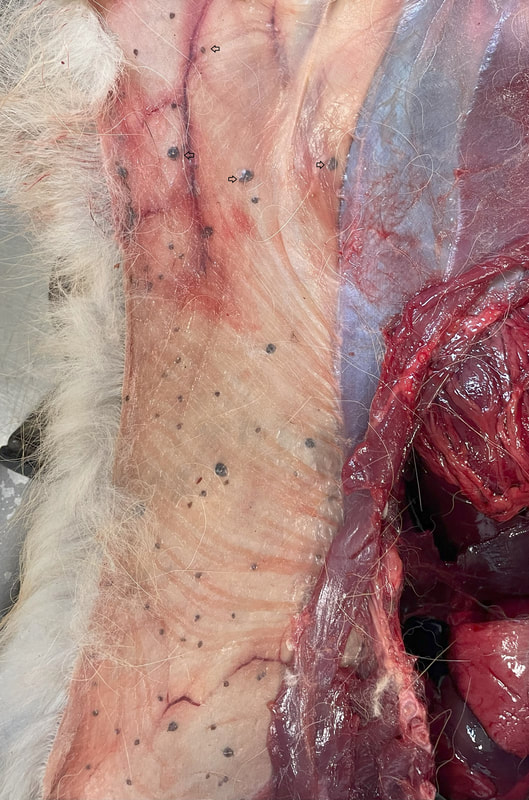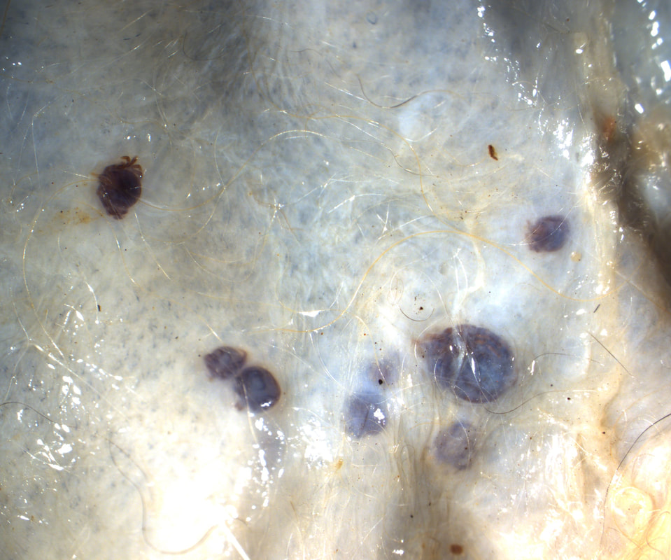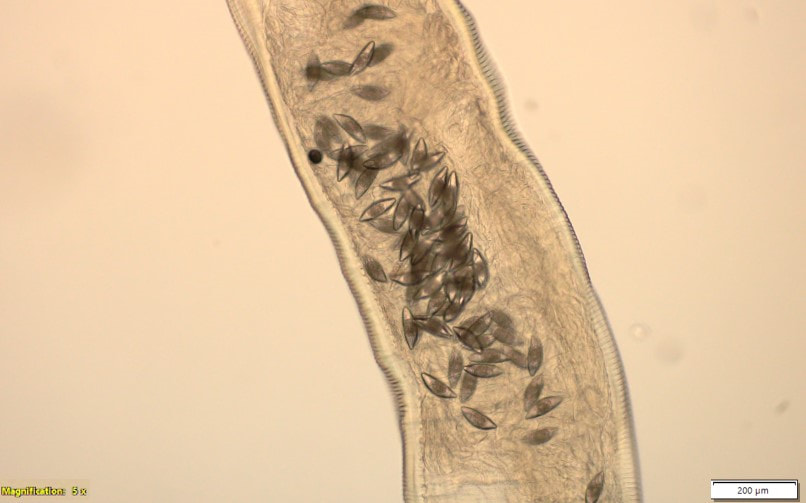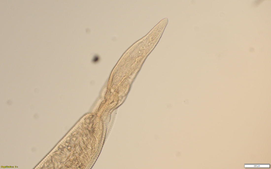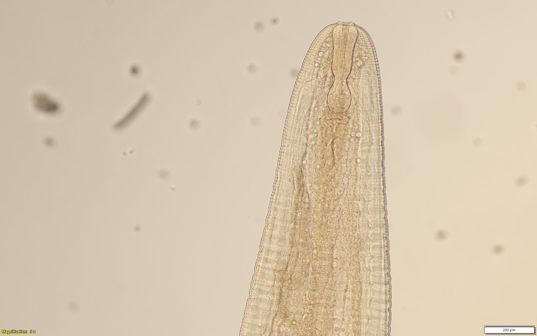
Not your usual case of formicationThe following images are from an approximately 2-year-old fox (Vulpes vulpes), necropsied at Oklahoma Animal Disease Diagnostic Laboratory. The fox was euthanized due to traumatic injury. This finding was incidental. (Images 1 and 2) Case contribution: Thank you to Ryan Carson-DVM class of 2026 and the AAVP student chapter at Oklahoma State University Image 1: Finding at necropsy Image 2: Microscopic view Subcutaneous Amblyomma americanum ticks (females, males, and nymphs). Ticks are obligate ectoparasites that spend part of their life attached to their hosts. They have mouthparts with chelicerae that pierce through the skin of the host. Attachment is facilitated by the tubular hypostome and a secreted cement or latex-like compound that attaches the tick to the host until the feeding is complete. Although most of the subcutaneous cases of subcutaneous ticks have described Ixodes spp., previous reports from Missouri and Arkansas have described the finding of intradermal infestations of red foxes (Vulpes vulpes) with Amblyomma americanum ticks. Other descriptions from other countries have mainly been from foxes, golden jackals, domestic and racoon dogs; and a single report also described the finding in a human. The reasons to explain the unusual location on these host are not clear yet, indications seem to suggest that immune response of canids may play a role. Bull all out of luckA 5-year-old Angus bull, was presented to the Veterinary Teaching Hospital at Oklahoma State University, with lethargy, coughing and wheezing of 3 weeks of duration. The following is an image of structures incidentally found on its blood smear (highlighted by circle). Case contribution: Thank you to Drs. Alys Harshbarger and Jim Meinkoth at Oklahoma State University for contributing to this case. 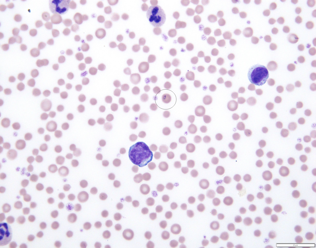 Theileria orientalis is a tick transmitted protozoan parasite that infects erythrocytes and leukocytes. In the United States, native genotypes are usually non-pathogenic, however in the past few years an increase of T. orientalis genotype Ikeda that causes diseases, has been observed. Clinical signs associated with this intracellular parasite are anemia, jaundice and weakness. Haemaphysalis spp. ticks are the primary biological vector of T. orientalis and are considered essential for completion of the lifecycle. This case was tested by PCR and DNA sequencing and detected as T. orientalis genotype chitose. Parasitic EmbraceA 3-month old calf was submitted for necropsy to the Oklahoma Animal Disease Diagnostic Laboratory. The calf was found lying down 2 days before dying. During necropsy examination, the body presented moderate emaciation and anemia. The cecal and colonic contents were pasty with numerous parasites 1 to 1.5 cm long. (Image 1) 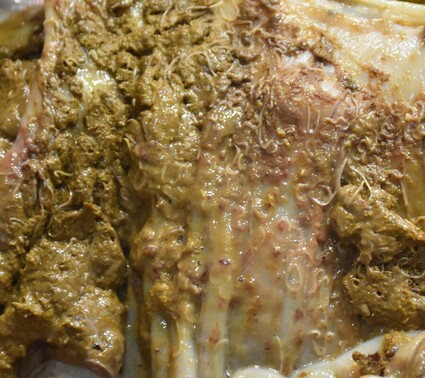 Image 1: Parasites found in cecal and colonic contents Oesophagostomum sp.
Oesophagostomins are parasites of the large intestine of ruminants, swine and primates. The adults are 1 to 2 cm long and the eggs are 70 to 90 x 34 to 45 um. They are also called nodular worms because their larva can become encapsulated by a reactive inflammation in hypersensitive hosts. Acute inflammation may lead to diarrhea that can be fatal. This calf also had high numbers of Moniezia sp. eggs, and Nematodirus helvetianus, with the last one being pathogenic in young animals that have no acquired immunity. (Image 2) 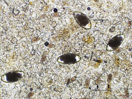
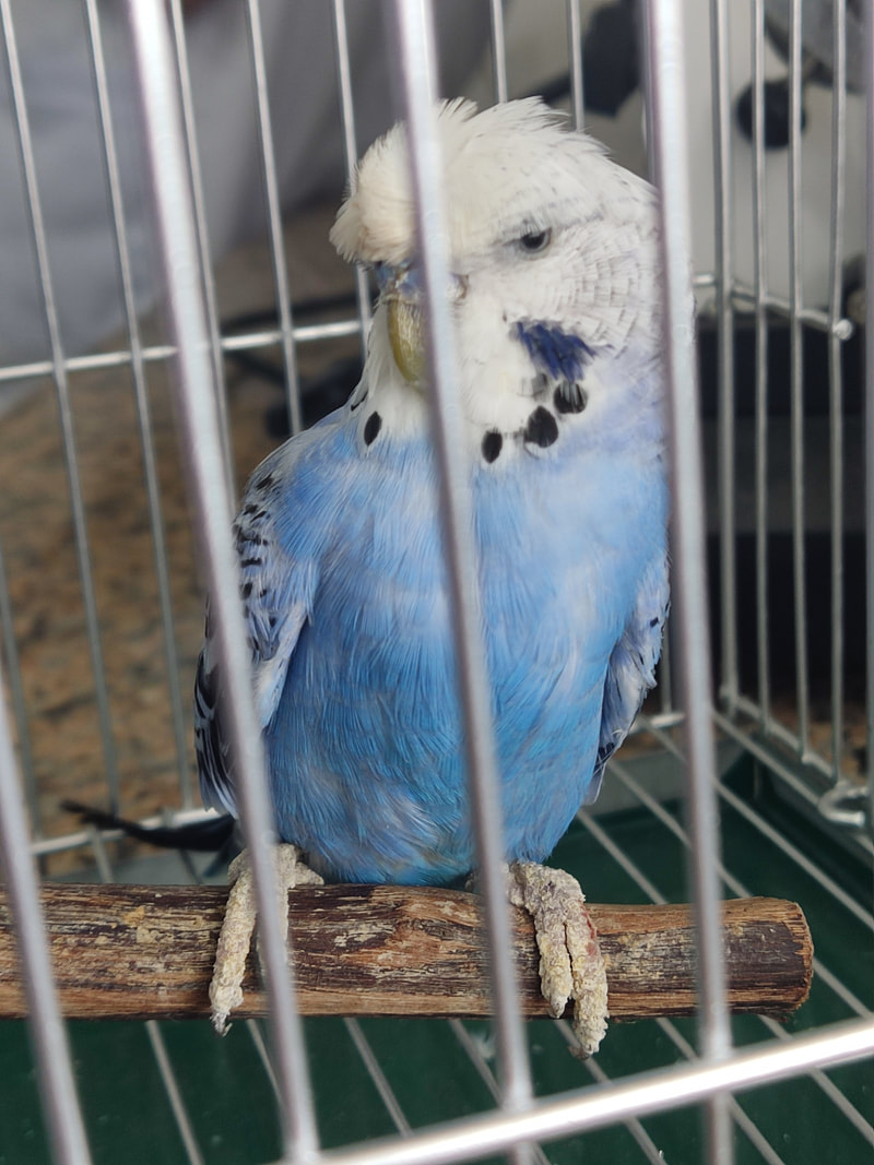
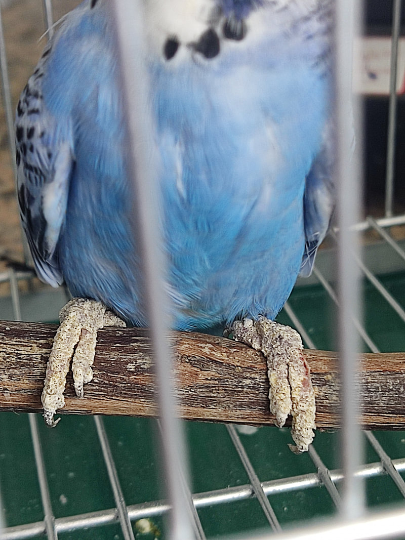
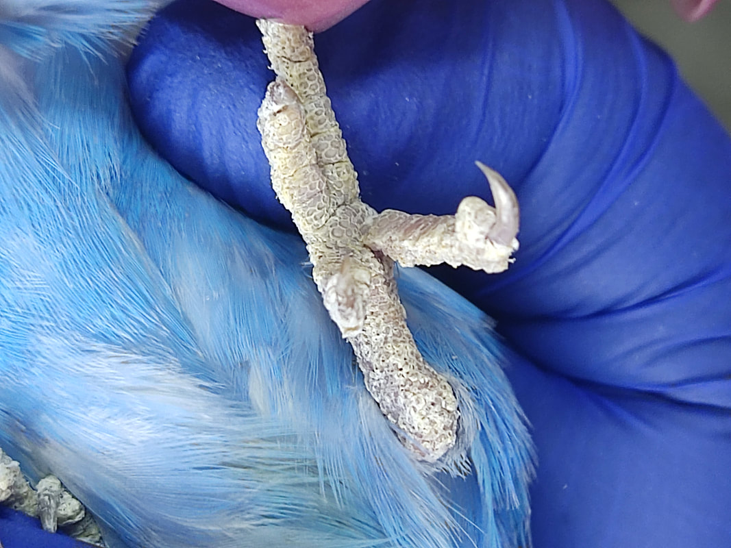
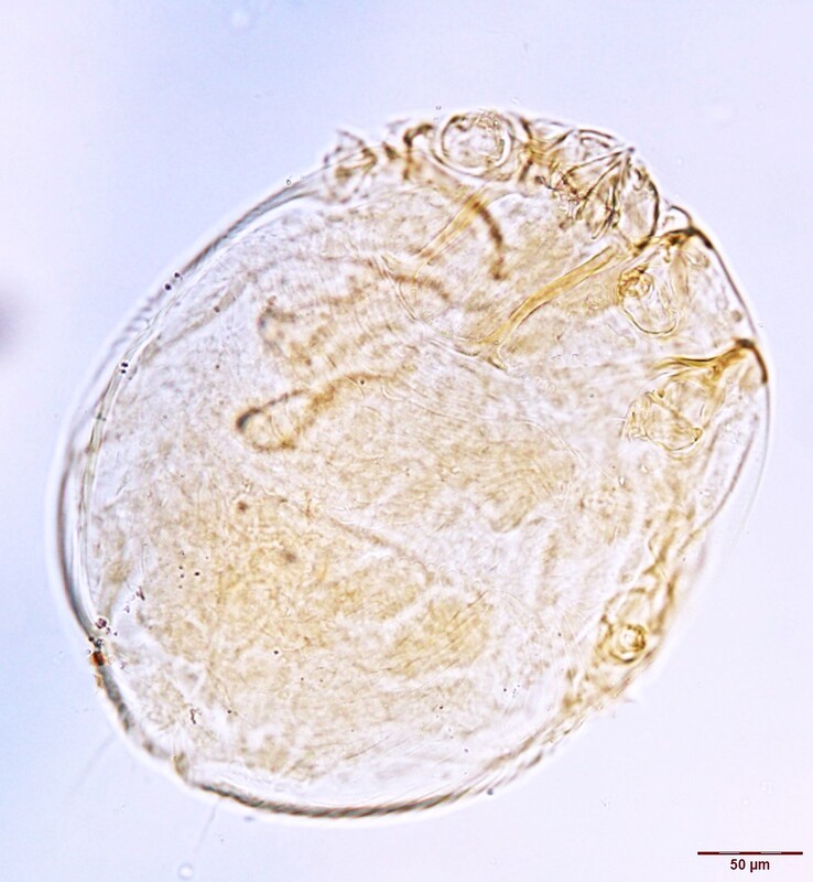
Image 2: Fecal centrifugation from small intestinal content Hope for a New Year's ResolutionA parakeet pet was presented to the College of Veterinary Medicine Clinic, Southwestern University in Philippines, with localized thickened, crusty lesions on the legs and feet, and scales lifting off on the area. No signs of pruritus were noticed. (Images 1-3) Microscopic examination of loose leg scales revealed the following. (Image 4) Case contribution: Erlah Shermaine Roble, DVM, Assistant Professor at the College of Veterinary Medicine. Southwestern University PHINMA. Philippines Images 1-3: Gross lesions upon presentation Image 4: Microscopic finding of leg scales Knemidocoptes sp. These mites burrow in the epidermis of the legs on birds, causing scales to lift and become loosened and the legs to become thickened and deformed. K. pilae is the species that commonly infest parakeets while K. mutans and K. jamaiciensis infest gallinaceous birds and canaries respectively. This case was resolved after oral administration of Ivermectin in drinking water. Christmas SurpriseA 2-year-old female Mongolian gerbil was submitted for necropsy to determine the cause of death as the owner has had multiple losses in the household. During necropsy, six specimens collected from the proximal area of the small intestine were incidentally found. All of the specimens recovered were identified as females containing spindle shaped eggs that measured 115 µm to 140 µm by 30 µm to 60 µm. (Images 1-3). Case provided by: Tiana Sanders DVM, PhD Student, Texas A&M Image 1: Eggs, 10x Image 2: Caudal end, 10x Image 3: Anterior end, 10x Dentostomella translucida. Known as the “Gerbil Pinworm” is a threadworm found in the small intestine of gerbils. Females typically measure 1 cm to 3 cm in length and males are 0.6 cm to 1 cm. The identifying features of D. translucida include a mouth with no lips, five unequal teeth per esophageal sector, and a thick, translucent, transversely striated cuticle. Typically, gerbils infected with D. translucida are asymptomatic and diagnosis may be difficult, as these parasites do not deposit their eggs around the perianal region. Worms on the BrainAn adult female woodchuck presented to a wildlife clinic in New York state after being found on the side of the road. The woodchuck had multifocal crusts and pustules over most of the body, with skin sloughing, with mites and fleas noted on external examination. The animal displayed neurologic signs, with, minimal response to handling, and paddling reported. A dewormer and antibiotics were administered, but despite treatment, the woodchuck continued to decline, with no interest in food by the third day. Euthanasia was elected, and the animal was submitted for necropsy following a negative rabies test. Histologic examination of the brain revealed the following: Case contribution: Timothy Wu, MS, DVM, Dipl. ACVP Image 1 and 2: Histologic findings in the brain Ascarid larva migrans (likely Baylisascaris sp.) Histologic evaluation reveals numerous up to 60 um diameter nematodes, determined to be the larval stage, given the absence of genital tracts within the examined sections. The presence of lateral alae (arrow), coelomyarian-polymyarian musculature, and a uninucleate intestine are consistent with ascarid larvae. While other ascarids, such as Toxocara canis cannot be ruled out, the most likely diagnosis in this case is considered to be Baylisascaris sp. There are many species of Baylisascaris affecting wildlife, including Baylisascaris columnaris in skunks, Baylisascaris transfuga in bears, and Baylisascaris laevis in woodchucks. The most commonly discussed species is Baylisascaris procyonis, which affects raccoons, and can cause visceral larva migrans in a wide range of hosts, including humans. Ascarid migration of the central nervous system, as in this case, can cause severe clinical signs. Although quick diagnosis and treatment with corticosteroids and albendazole can prove effective, this is not always plausible, as diagnosis can prove difficult. |